
- Home Page
- Company Profile
-
Our Products
- Microscope
- Stereo Inspection Scope
- Stereo Inspection Microscope RSMr-X8
- Polarizing Projection Microscope RPL-4
- Portable Metallurgical Microscope RMM-5A
- Inverted Metallurgical Microscope RMM-77
- Advanced Research Microscope RXLr-5NXM2
- Confocal Microscope RTC-7 CON
- Trinocular Upright Metallurgical Microscope RXM-7T
- Senior Dissecting Microscope RDM-4
- Inverted Fluorescence Microscope RTC-7A
- Metallurgical Microscope RMM-88
- Trinocular Coaxial Microscope RXL-4T
- Senior Inspection Spinneretscope RIS-30
- Biological LCD Microscope RXL-4LCD
- CONFOCAL MICROSCOPES RTC-7 CON
- Portable Grooved Metallurgical Microscope RMM-6L
- Digital Spinneret Inspection Microscope RIS-45
- Toolmaker Microscope Large RTM-900DR
- Dissecting Microscope RDM-2
- Advance Stereo Zoom Microscope RSMr-10
- Toolmaker's Microscope RTM-900DL
- Biological Research Microscope RXLr-5 Series
- Trinocular Stereo Zoom Microscope RSM-9
- Motorized Comparision RCM-18
- Digital Research Microscope RXLr-4NX
- Deca Head Microscope RXLr-5 Nx
- BINOCULAR STEREO ZOOM MICROSCOPE RSM-8AS
- Trinocular Projection Microscope PRM-18T
- SPINNERET MICROSCOPE RIS-90
- Binocular Research Microscope RXL-5B
- Trinocular Polarizing Microscope RPL-55T
- Penta Head Microscope/ Multi View Head Microscope RXLr-5000
- Sieves Digital Microscope RSP-90
- PCB Inspection Video Stereoscope Microscope RPCB-45
- Trinocular Inverted Tissue Culture Microscope RTC-8A
- Tool Maker's Microscope RTM-500
- Stereo Microscope RSM-4
- Student School Microscope RM-2
- Student Compound Microscope RM-2A
- Student Projection Microscope PRM-11A
- Portable Inverted Tissue Culture Microscoe RTC-1P
- Upright Binocular Research Microscope RXLr-5Nx
- Projection Microscope PRM-12/12A
- Gemological Microscope RGM-8
- Digital Biological Microscope RXLr-4D
- Student Medical Microscope RM-3
- Inverted Tissue Culture Microscope RTC-6
- Laboratory Microscope RM-600B
- Advanced Research Material Microscope RXLr-5M
- Coaxial Microscope RXL-4
- Tissue Culture Microscope RTC-5
- Advanced Inverted Phase Microscope RTC-7NX
- Measuring Microscope ROM-100D
- RXL-5 DIGI 40X-600X
- Digital Biological Microscope 40X-1600X
- Microtomes
- Hand Table Microtome RMT-5
- Rotary Microtome RMT-20A
- Slide Staining Machine RSSD-23
- Senior Precision Rotary Microtome RMT-30
- Knife Sharpener Microtome RSM-100
- Advance Rotary Microtome RMT-30A
- Freezing Microtome RMT-40
- Rotary Microtome RMT-20 (Erma Type)
- Rocking Microtome RMT-10
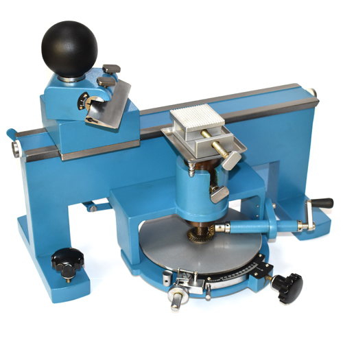
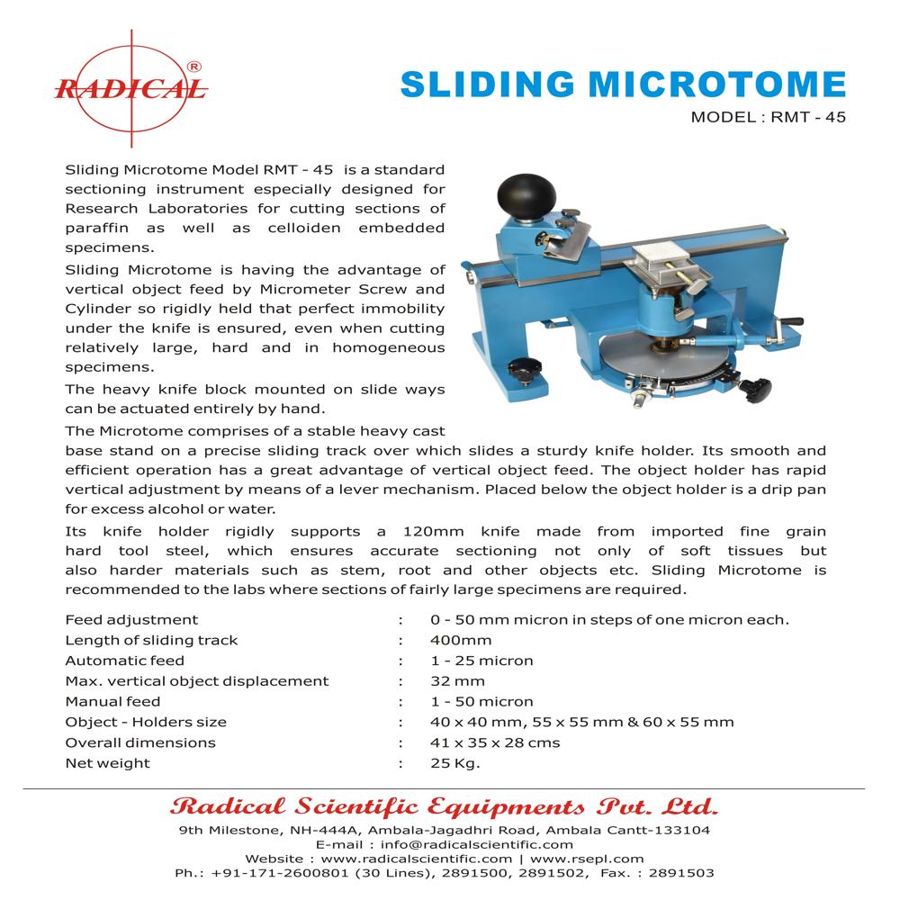
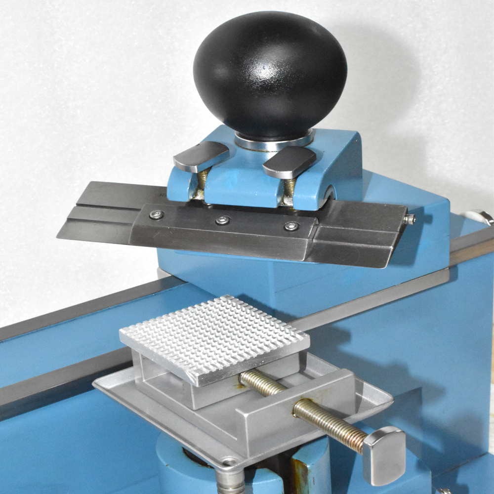
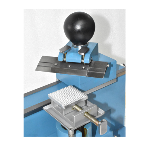
- Sliding Microtome RMT-45
- Manual Rotary Microtome RMT-25
- Fully Automatic Rotary Microtome RMT-75
- Precision Fiber Microtome RMT-55
- CRYOSTAT MICROTOME RSC-28
- Optical Instruments
- Optical Charpy Projector RPP-250C
- Profile Projector RPP-3000DP
- Crack Width Ruler RWR-7
- Digital Vertical Autoclave RAV-50
- Universal Profile Projector RPP-3000
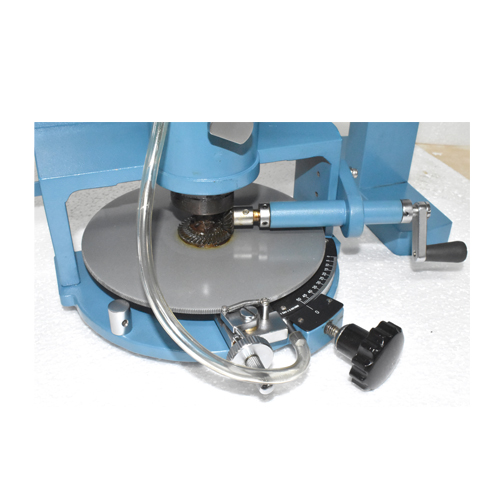
- Automatic Metallographic Polishing Machine RSMP-2S
- Illuminated Flexible Arm Magnifier RBM-104
- Gem Illuminated Spectroscope RDV-77
- ILLUMINATED OPTICAL COMPARATOR RBM-103
- Shop Microscope (Measuring Microscope) ROM Series
- Brinell Microscope RBM-55
- Grooved Laboratory Microscope RMM-6L
- Bench Magnifier RBM-101
- Flexible Arm Magnifier RBM-104L
- Table Top Magnifier Aluminum RBM-105
- POLARISCOPE Model : RSPS-24
- Tissue Processor Machine
- Microscope Accessories
- Histopathological Equipment
- SEMI AUTOMATIC ROTARY MICROTOME RMT-35
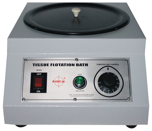
- Tissue Flotation Water Bath RSTI-138
- DIGITAL LABORATORY INCUBATOR RSTI-Series
- Laboratory Incubator RSTI-108
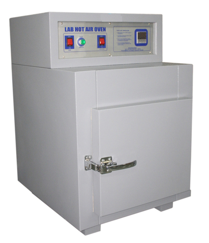
- Hot Air Oven Digital RSTI-102
- Bone and Brain Cutting Machine RBCM-55
- Embalming Machine RSTI-130
- SLIDING MICROTOME RMT-45
- High Temperature Oven RSTI-108
- Laminar Air Flow Horizontal
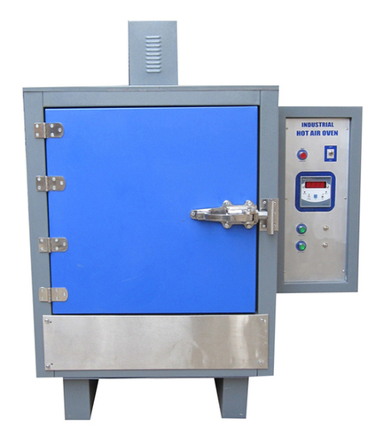
- Industrial Hot Air Oven RSTI-104
- Slide Cabinet RS-105 Series
- Vertical Autoclave
- Flocculator Jar Test Apparatus RS-1924
- Lab Instrument
- BOD INCUBATOR RSTI-110
- Micro Centrifuge 16000 R.P.M. RST-16
- Dissolved Oxygen Meter RS-801
- Incubator Shaker Water Bath RSTI-140
- Hemoglobin Meter (Sahli's) RSHB-10
- Water Distillation with Metal Heater RSLWS-MH-40M
- Tablet Dissolution Test Apparatus (6 Basket) RS-1916
- Precision Water Bath RSTI-137
- Digital Magnetic Stirrer RSTI-156
- GROSSING TABLE
- Disintegration Test Apparatus RS-901
- Digital Photo Colorimeter RSPC-9
- Double Disc Polishing Machine Semi Automatic RSMP-2S+MPT
- Laboratory Rectangular Hot Plates RSTI-146
- Vortex Shaker RSTI-151
- MAGNETIC STIRRER with HOT PLATE RSTI-156
- Haemoglobin Meter RSHB-50
- Dual Channel Flame Photometer RS-391
- Round Centrifuge RSC-15
- pH or mv or Conductivity
- VDRL ROTATOR (VARIABLE SPEED) RSTI-150
- Haematocrit Centrifuge RST-15
- Rectangular Water Bath RSTI-135
- PASS BOX DYNAMIC
- Serological Water Bath RSTI-134
- Digital Turbidity Meter RS-335
- Heating Mantle RSTI-148
- Revolutionary General Purpose Digital Centrifuge RST-8M
- Hand Specimen Leveler Press MM-18
- COOLING PLATE
- MICROPROCESSOR PH METER RS-1013
- DIGITAL FLAME PHOTOMETER RS-1381
- pH/mV/TEMPERATURE TESTER RS-7011
- DIGITAL PH, CONDUCTIVITY & TEMPERATURE METER RS-101
- Auto Karl Fischer Titrimeter RT-761
- Atomic Model Set (Euro Design)
- Laboratory DEEP Freezer RSTI-121
- Blood Bank Refrigerator RSTI-126
- Heating Mantles RSTI-148
- Laboratory Round Hot Plate RSTI-145
- Melting Point Apparatus RS-934
- Jominy End Quench Apparatus RJQA-45
- Digital PH Meter
- DELUX PH METER-101
- BULK DENSITY APP - 951
- Microprocessor PH Meter - 1015
- Microprocessor Colony Counter 1361
- Microprocessor colony counter 1362
- Microprocessor Colony Counter RS-1363
- Tap Density Tester RS-1951
- Alpha03 Digital Ph Colorimeter
- DIGITAL PH METER (PORTABLE) RS-131& 132
- PH/MV/TEMPERATURE- 7200
- Stereo Zoom Microscope
- Universal Stereo Zoom Microscope RSM-8U
- Jewellery Making Microscope RGM-10
- Motorized Stereo Microscope RSMr-X8
- Advance Stereo Zoom Microscope RSMr-3T
- Stereo Inspection Microscope RSM-15
- Stereo Zoom Microscope RSM-8
- Stereo Microscope RSM-4T
- Digital 3D Inspection Microscope RSZ-3D
- Articulated Trinocular Stereo Zoom Microscope RSM-9AS
- Binocular Stereo Microscope RSM-5
- Binocular Stereo Microscope RSM-4F
- Digital 3D Inspection Microscope RSZ-3D
- Polarising Microscopes
- Advanced Polarizing Microscope RPL-3B
- Binocular Polarizing Microscope RPL-55B
- Laboratory Polarizing Microscope RPL-1
- Trinocular Polarising Microscope RPL-3T
- Advanced Research Polarizing Microscope RXLr-4T
- BINOCULAR RESEARCH POLARIZING MICROSCOPE RPL-55B
- LABORATORY POLARIZING MICROSCOPE RPL-1
- Polarizing Microscope RPL-3T
- Advanced Polarizing Microscope RPL-3B
- Lab Consumables
- Carboy with Wide Handle
- ORBITAL SHAKING INCUBATOR RSTI-111
- Atomic Model Set
- Slide Mailer
- Desiccator (Vaccum)
- Desiccator (plain)
- Carboy with Stopcock
- Atomic Model Set (PP)
- DIGITAL LABORATORY RECTANGULAR HOT PLATES RSTI-146
- Scoop PP
- Dropping Bottles
- Wash Bottles
- Coplin Jar Polypropylene
- Slide Box
- Slide Tray
- Slide Draining Tray
- PCR Tube Rack with Hinges - Autoclavable
- MCT Box
- Rotatable Multi Rack
- Measuring Cylinder Hexagonal
- Draining Lab Rack
- Photo Colorimeter- 1311
- Profile Projector
- PROFILE PROJECTOR
- Charpy Profile Projector RPP-250C
- Compact, Table Top Profile Projector
- High Sharpness Profile Projector RPP-350 DR
- Profile Projector Shadowgraph RPP-3000DP
- Vertical Profile Projector RPP-60DR+
- Coaxial Profile Projector RPP-3000
- Profile Projector RPP-350
- Bench Type Profile Projector RPP-500HDR
- Profile Projector RPP-500
- Profile Projector RPP-150
- Profile Projector (Floor Model) RPP-60DR
- Anotomy Model
- Metallurgical Microscope
- INVERTED METALLURGICAL MICROSCOPE RMM-88S
- Advanced Laboratory Metallurgical Microscope RXLr-4M
- Research Material Microscope
- Emery Paper (Velvet)/Sand Paper
- Belt Grinder/Polisher RBP-100
- Single Disc Polishing Machine RPM-22S
- Double Disc Digital Polishing Machine RPM-33D
- ABRASIVE CUT-OFF MACHINE RACM-55
- Metallographic Specimen Mounting Press RHM-2B-30
- Metallographic Hot Mounting Machine RHM-5
- Digital Hot Mounting Machine RHM-2
- Portable Metallurgical Microscope RMM-5L
- Lab items
- Microscope
- Contact Us


 Call Me Free
